In stockBestseller
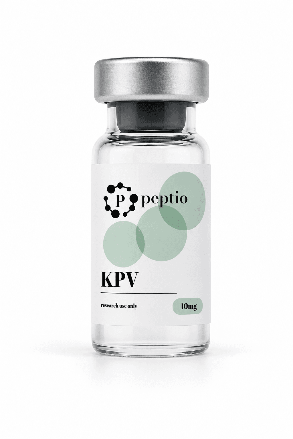
For research use only · Not for human consumption
KPV is the last three residues of α-melanocyte-stimulating hormone: lysine, proline and valine. The parent hormone is a tridecapeptide derived from proopiomelanocortin, written SYSMEHFRWGKPV, and the tripeptide is its C-terminal end (1)(3).
Hiltz and Lipton tested that fragment on its own in 1989, giving graded doses to mice and comparing the effect on picryl chloride-induced ear swelling against saline and against a large dose of corticosteroid. The fragment inhibited swelling in a dose-related fashion, and that is where the idea that this end of the molecule carries general anti-inflammatory activity comes from (2).
Almost everything published since is cell culture and rodent work. The studies cited on this page used human intestinal epithelial cell lines and human T cells in culture, mouse models of colitis, mouse peritonitis, and — in the one study that used human tissue — excised skin rather than a person. There is no controlled human trial of the tripeptide, and a ClinicalTrials.gov search for KPV as an intervention returned no registered studies when this page was written.
So this page publishes no figures. There is no percentage change to report, no dose-response chart and no placebo arm, because no trial has produced any. What follows describes the mechanisms the published work proposes, and the models it proposed them in.
α-MSH is itself a well-described anti-inflammatory peptide, and it is also the hormone that darkens skin. Separating those two activities is the whole reason a fragment of three residues became interesting, and the 2008 Endocrine Reviews survey of the field states the position directly: the pigmentary effect is the obstacle limiting the use of α-MSH in inflammatory disorders, and the C-terminal tripeptide has been put forward as an alternative because it preserves the anti-inflammatory effect while lacking the pigmentary action (3).
The structural reason is that the melanocortin message sits elsewhere in the molecule. His-Phe-Arg-Trp — the core melanocortin sequence — lies in the middle of the parent hormone and is not part of the C-terminal tripeptide (1)(5). Getting and colleagues compared the two in a mouse peritonitis model and found the tripeptide behaving unlike the core peptides in three ways: it did not raise cAMP in macrophages, its antimigratory effect was not blocked by the MC3/4-R antagonist SHU9119, and it stayed active in recessive yellow mice, whose MC1R is non-functional. They concluded that it is unlikely to act through melanocortin receptors at all, and more likely through inhibition of interleukin-1β functions (5).
That is the most interesting thing about this molecule, and it is also entirely a statement about mice and cultured cells. None of the cited work establishes what either peptide does in a person.
NF-κB
Transcription factor and MAP kinase signalling
Dalmasso and colleagues stimulated human intestinal epithelial cell lines — Caco2-BBE and HT29-Cl.19A — and Jurkat T cells with pro-inflammatory cytokines, then read the response with an NF-κB luciferase reporter, Western blot, real-time RT-PCR and ELISA. Nanomolar concentrations of the tripeptide inhibited activation of NF-κB and of MAP kinase signalling and reduced pro-inflammatory cytokine secretion (4).
MC1R
Melanocortin receptors, and doing without them
Two groups tested whether melanocortin receptors are required, and both reported that they are not, at least not fully. In crystal-induced peritonitis the effect was not blocked by an MC3/4-R antagonist and persisted in mice with a non-functional MC1R (5). In DSS colitis, Kannengiesser and colleagues reported effects that appear at least partially independent of MC1R signalling, and their MC1R-deficient treatment group was rescued from death (6). The peritonitis work also records a negative result: unlike α-MSH, the tripeptide did not inhibit macrophage activation in culture (5).
PepT1
Transport into the epithelium
PepT1 is a di- and tripeptide transporter normally expressed in the small intestine and induced in the colon during inflammatory bowel disease. Uptake experiments using radiolabelled tripeptide, and competition against a known PepT1 substrate, indicated that the peptide enters intestinal epithelial and immune cells through that transporter — the explanation offered for how a peptide given in drinking water reached inflamed tissue in the mouse studies (4).
Two papers from 2008 carry most of the in vivo work, and both are mouse colitis studies. Dalmasso and colleagues added the tripeptide to drinking water and reported a reduced incidence of colitis induced by dextran sulfate sodium and by trinitrobenzene sulfonic acid, assessed histologically and by pro-inflammatory cytokine mRNA expression (4). Kannengiesser and colleagues used DSS colitis and a CD45RBhi transfer model, measuring body weight, colon histology and myeloperoxidase activity, and reported significant anti-inflammatory effects in both (6).
The delivery literature is newer and equally preclinical. Xiao and colleagues loaded the tripeptide into polymeric nanoparticles functionalised with hyaluronic acid, gave them orally inside a chitosan and alginate hydrogel, and reported in a mouse ulcerative colitis model that the carrier both accelerated mucosal healing and lowered TNF-α (7).
Pawar and colleagues are the one group in this set to have worked on human tissue, and it was excised skin on a diffusion cell rather than a participant. Passive permeation across intact skin fell below the assay's detection limit; the peptide crossed only after the skin was microporated, and more again with iontophoresis (8). What that establishes is a delivery problem, not an effect.
One older finding sits outside inflammation altogether. Cutuli and colleagues reported that α-MSH and its C-terminal tripeptide inhibited colony formation by Staphylococcus aureus and reduced viability and germ tube formation in Candida albicans, across a concentration range extending down to the picomolar, and attributed it to raised cellular cAMP (1).
Three peptides that share a shelf and not much else. No cell in this table holds a figure, because none of the three has a published controlled trial in people to take one from. The rows report what the cited work says about where each peptide comes from, the pathway it proposes, the models it used and the route it used — none of it is a comparison of effect, and no study compared the three against each other.
| Parameter | KPV | BPC-157 | TB-500 |
|---|---|---|---|
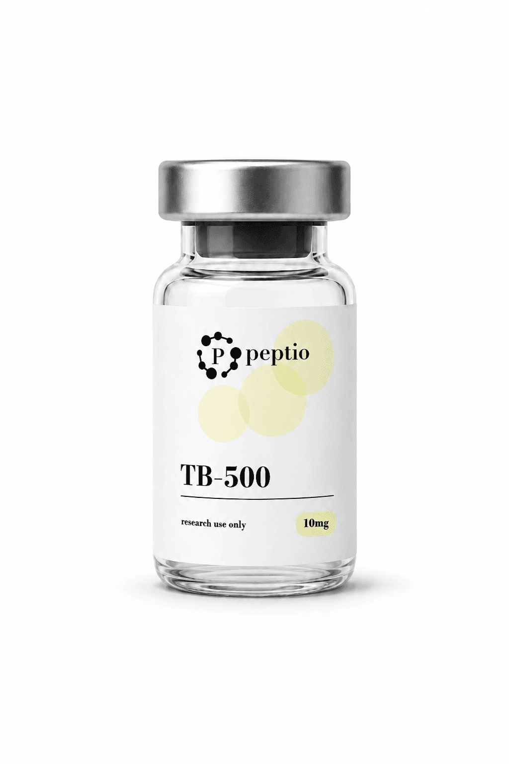
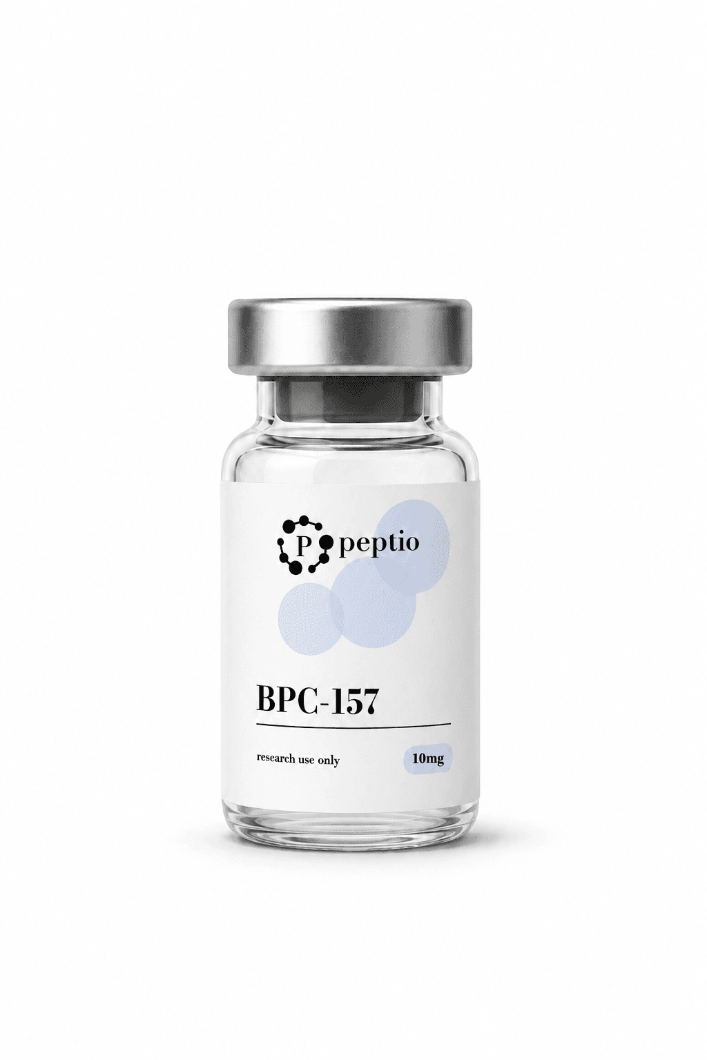
| Origin | C-terminal tripeptide of α-melanocyte-stimulating hormone (1)(5) | Pentadecapeptide, a partial sequence of a protective compound described in human gastric juice (9) | Synthetic version of thymosin β-4, an actin-sequestering protein of eukaryotic cells (10) |
| Pathway proposed in the cited work | Inhibition of NF-κB and MAP kinase signalling, with reduced pro-inflammatory cytokine secretion (4) | The nitric oxide system and angiogenic growth factor signalling, including a VEGFR2–Akt–eNOS route (9) | Actin sequestration, with a described role in dermal and corneal wound repair (10) |
| Relationship to a receptor | Effect retained without a functional MC1R and not blocked by an MC3/4-R antagonist (5)(6) | No single receptor identified in the review cited here (9) | Described as acting on intracellular actin rather than a named surface receptor (10) |
| Evidence stage in the sources cited here | Cell culture and mouse colitis models (4)(6) | Rat, mouse and pig injury models (9) | Cell models and a rat full-thickness wound model (10)(11) |
| Route used in those studies | Oral in drinking water (4); oral in nanoparticle carriers (7); systemic injection in peritonitis (5) | Oral, intraperitoneal, topical and local application at the injury (9) | Topical and intraperitoneal in the rat wound model (11) |
| Marketing authorisation | None, in any jurisdiction | None, in any jurisdiction | None, in any jurisdiction |
Sources. (1) Cutuli M, Cristiani S, Lipton JM, Catania A. Antimicrobial effects of alpha-MSH peptides. J Leukoc Biol. 2000;67(2):233–239. (2) Hiltz ME, Lipton JM. Antiinflammatory activity of a COOH-terminal fragment of the neuropeptide alpha-MSH. FASEB J. 1989;3(11):2282–2284. (3) Brzoska T, Luger TA, Maaser C, Abels C, Böhm M. Alpha-melanocyte-stimulating hormone and related tripeptides: biochemistry, antiinflammatory and protective effects in vitro and in vivo, and future perspectives for the treatment of immune-mediated inflammatory diseases. Endocr Rev. 2008;29(5):581–602. (4) Dalmasso G, Charrier-Hisamuddin L, Nguyen HTT, Yan Y, Sitaraman S, Merlin D. PepT1-mediated tripeptide KPV uptake reduces intestinal inflammation. Gastroenterology. 2008;134(1):166–178. (5) Getting SJ, Schiöth HB, Perretti M. Dissection of the anti-inflammatory effect of the core and C-terminal (KPV) alpha-melanocyte-stimulating hormone peptides. J Pharmacol Exp Ther. 2003;306(2):631–637. (6) Kannengiesser K, Maaser C, Heidemann J, et al. Melanocortin-derived tripeptide KPV has anti-inflammatory potential in murine models of inflammatory bowel disease. Inflamm Bowel Dis. 2008;14(3):324–331. (7) Xiao B, Xu Z, Viennois E, et al. Orally targeted delivery of tripeptide KPV via hyaluronic acid-functionalized nanoparticles efficiently alleviates ulcerative colitis. Mol Ther. 2017;25(7):1628–1640. (8) Pawar K, Kolli CS, Rangari VK, Babu RJ. Transdermal iontophoretic delivery of lysine-proline-valine (KPV) peptide across microporated human skin. J Pharm Sci. 2017;106(7):1814–1820. (9) Seiwerth S, Milavic M, Vukojevic J, et al. Stable gastric pentadecapeptide BPC 157 and wound healing. Front Pharmacol. 2021;12:627533. (10) Goldstein AL, Hannappel E, Kleinman HK. Thymosin beta4: actin-sequestering protein moonlights to repair injured tissues. Trends Mol Med. 2005;11(9):421–429. (11) Malinda KM, Sidhu GS, Mani H, et al. Thymosin beta4 accelerates wound healing. J Invest Dermatol. 1999;113(3):364–368. No cell in this table reports a clinical figure, because no controlled human trial of any of the three has been published. KPV holds no marketing authorisation in any jurisdiction.
A sealed vial of lyophilised powder, in a 5 mg or a 10 mg size. Both are the same material; the difference is the mass in the vial, not the concentration — concentration is set later, by how much solvent is added.
Each batch is released against a certificate of analysis from an independent issuer, and the batch number printed on the vial matches the number on that certificate. Checking those two against each other is the most useful thing to do with a delivery. The purity, form, storage condition and molecular weight recorded for the current batch are in the specifications above.
Handling is the same as for every other compound in the catalogue. The handling protocol below covers storage temperatures, solvent choice and the concentration calculation.
Research use only
KPV is not an approved medicine and holds no marketing authorisation in any jurisdiction. Peptio is not a pharmacy and supplies it for research use only — not for human or veterinary consumption. Everything reported on this page comes from published cell culture and animal studies and describes those models; none of it is guidance for use in people, and no controlled human trial of this tripeptide has been published.
Every compound Peptio ships arrives as a sealed vial of lyophilised powder. Freeze-drying is what keeps it stable in transit: with the water removed and the vial closed, the peptide keeps far longer than it would in solution. The protocol below applies to all of them, and it is the same protocol whichever compound is in the vial.
The steps that matter most happen in the first few minutes after a vial is opened, and in how the prepared solution is labelled afterwards. A vial with no recorded solvent volume is a vial with no known concentration, and no amount of care later recovers that.
01
Inspect on arrival
Check that the crimp seal and stopper are intact and that the powder cake is dry and unbroken. Compare the batch number printed on the vial with the number on the certificate of analysis. A cake that has slumped into a film, or a vial that arrives loose in its packaging, is worth photographing before anything else is done to it.
02
Equilibrate before opening
Let a vial taken from cold storage stand sealed until it reaches room temperature. Opening cold glass in a warm room pulls condensation onto it, and moisture is the one thing a desiccated powder is being protected from.
03
Add the solvent
Direct the solvent slowly down the inner wall of the vial rather than onto the cake, then let it dissolve on its own and swirl gently until the solution runs clear. This takes a minute or two, and it is not helped by shaking.
04
Label, then refrigerate
Write the date, the solvent and the exact volume added onto the vial before it goes anywhere. Keep the prepared solution cold and out of direct light, and check that it is still clear each time it is used.
| Correct | Common mistake |
|---|---|
| Direct the solvent down the inner wall of the vial and let the cake dissolve on its own. | Drive the solvent onto the powder in a fast stream. |
| Swirl gently until the solution runs clear. | Shake or vortex the vial — mechanical agitation and foaming degrade peptide structure. |
| Let a vial from cold storage reach room temperature while it is still sealed. | Open a vial straight out of the freezer, and pull condensation into a product that is kept dry on purpose. |
| Label every prepared vial with the date, the solvent and the volume added. | Rely on memory — two vials that look identical at 2–8 °C can hold two different concentrations. |
| Draw through the stopper with a fresh sterile needle each time. | Prise off the crimp seal; an open vial cannot be kept sterile. |
| Keep prepared solution cold and shielded from light between uses. | Leave a reconstituted vial standing at room temperature on the bench. |
| Look at the solution before every use and confirm it is clear. | Use a solution that has turned cloudy or thrown a visible particle. |
Solvent volume sets the concentration
The mass of peptide in a vial is fixed at manufacture. The only variable is how much solvent goes in, and it changes every measurement that follows — 2 mL instead of 1 mL halves the concentration. Confirm the vial's usable capacity before adding anything, since a 2 mL vial will not take 3 mL, and write down the volume you actually added.
Questions
0.0
0 reviews
No reviews yet.